
Raman scattering is named after Sir Chandrasekhara Venkata Raman who discovered the phenomenon in 1928. Raman scattering is a type of inelastic light scattering. This means that light interacts with a molecule and changes its energy through that interaction. In the case of Raman scattering, the light excites vibrations in the scattering molecule. These vibrations require energy, so the scattered light has less energy.
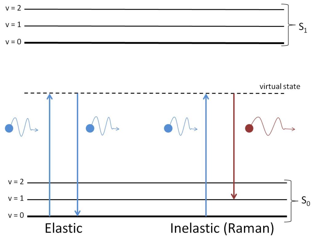
Every molecule will vibrate differently. This means that each type of molecule will scatter light with different changes of energy. The different energies of scattered light act as a “molecular fingerprint” we can use to identify the types and quantities of molecule in a sample.
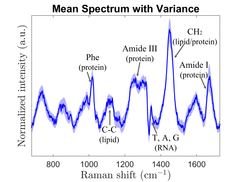
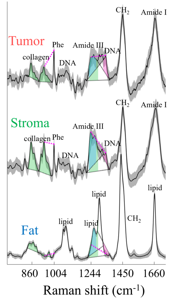
This molecular specificity makes Raman spectroscopy a powerful tool for chemical analysis. Raman spectroscopy can find slight differences in the molecular content of tumor vs. healthy cells, different species of bacteria, or numerous other samples.
A basic Raman system consists of a laser, a spectrometer, and a detector. The laser sends a single wavelength to excite molecular vibrations in the sample. Each energy shift due to these vibrations results in light scattered a different wavelength. These wavelengths are separated by the spectrometer and measured by a very sensitive detector. Raman scattering events are not common (about one Raman scattered photon for every million incident photons), so even with a sensitive detector, each Raman measurement can take several seconds or even minutes.
For more detailed information on Raman spectroscopy, see these reviews:
- Durrant, B., Trappett, M., Shipp, D., & Notingher, I. (2019). Recent developments in spontaneous Raman imaging of living biological cells. Current opinion in chemical biology, 51, 138-145.
- Shipp, D. W., Sinjab, F., & Notingher, I. (2017). Raman spectroscopy: techniques and applications in the life sciences. Advances in Optics and Photonics, 9(2), 315-428.
